
Vol.:(0123456789)
1 3
Journal of Neurology
https://doi.org/10.1007/s00415-020-10001-7
LETTER TOTHEEDITORS
Reversible Encephalopathy Syndrome (PRES) inaCOVID‑19 patient
LuciaPrinciottaCariddi
1,5
· PayamTabaeeDamavandi
1,6
· FedericoCarimati
1
· PaolaBan
1
· AlessandroClemenzi
1
·
MargheritaMarelli
2
· AndreaGiorgianni
3
· GabrieleVinacci
4,5
· MarcoMauri
1,7
· MaurizioVersino
1,7
Received: 3 June 2020 / Revised: 13 June 2020 / Accepted: 16 June 2020
© Springer-Verlag GmbH Germany, part of Springer Nature 2020
Abstract
Recently WHO has declared novel coronavirus disease 2019 (COVID-19) outbreak a pandemic. Acute respiratory syndrome
seems to be the most common manifestation of COVID-19. Besides pneumonia, it has been demonstrated that SARS-CoV-2
infection affects multiple organs, including brain tissues, causing different neurological manifestations, especially acute
cerebrovascular disease (ischemic and hemorrhagic stroke), impaired consciousness and skeletal muscle injury. To our
knowledge, among neurological disorders associated with SARS-CoV2 infection, no Posterior Reversible Encephalopathy
Syndrome (PRES) has been described yet. Herein, we report a case of a 64-year old woman with COVID19 infection who
developed a PRES, and we suggest that it could be explained by the disruption of the blood brain barrier induced by the
cerebrovascular endothelial dysfunction caused by SARS-CoV-2.
Keywords Reversible encephalopathy syndrome PRES· COVID-19· Endothelialdysfunction
Abbreviations
COVID-19 Corona virus disease 19
CTA Computed tomography angiography
ED Endothelial dysfunction
SARS-Cov2 Severe acute respiratory syndrome covid 2
Case presentation
A 64-year-old woman was admitted to our hospital with a
10-day history of fever and dyspnea treated at home with
ceftriaxone.
Her medical history included hypertension, gastroesopha-
geal reflux disease, hyperuricemia, dyslipidemia, obstructive
sleep apnea and paroxysmal atrial fibrillation. Her medica-
tions were: irbesartan/hydrochlorothiazide, acetylsalicylic
acid, pantoprazole, rosuvastatin, allopurinol and bisoprolol.
She was febrile (39°C) with marked dyspnea. Neurologi-
cal examination was unremarkable. Laboratory tests were
significant for lymphocytopenia with increased transami-
nases and LDH. Oxygen saturation was low, thereby oxygen
therapy was administered (Table1). Chest X-ray showed
reduction of the parenchymal transparency in basal region
of right lung.
A continuous positive airway pressure had to be started.
A nasopharyngeal swab resulted positive for SARS-CoV-2;
antiviral therapy with darunavir/cobicistat, associated with
hydroxychloroquine were started. After 24h, she was taken
to Intensive Care Unit: she was sedated and mechanical
ventilation was started. Antiviral plus antibiotic therapies
were continued for 10days. After 23days bronchial aspirate
turned negative for SARS-CoV-2.
On day 25 she woke up when sedation was weaned; she
was drowsy and complained of blurred vision. She showed
Lucia Princiotta Cariddi and Payam Tabaee Damavandi
contributed equally as first authors.
Marco Mauri and Maurizio Versino contributed equally as last
authors.
* Maurizio Versino
1
Neurology andStroke Unit, ASST Sette Laghi, Circolo
Hospital, Viale Borri, 57, 20100Varese, Italy
2
Pneumology Unit, ASST Sette Laghi, Circolo Hospital,
Varese, Italy
3
Neuroradiology Unit, ASST Sette Laghi, Circolo Hospital,
Varese, Italy
4
Radiology Unit, ASST Sette Laghi, Circolo Hospital, Varese,
Italy
5
Clinical andExperimental Medicine andMedical
Humanities, Center ofResearch inMedical Pharmacology,
University ofInsubria, Varese, Italy
6
University ofMilano Bicocca, Monza, Italy
7
University ofInsubria, Varese, Italy

Journal of Neurology
1 3
an altered mental status, a decreased left nasolabial fold, the
tone and the strength were slightly decreased in the legs, and
all deep tendon reflexes were reduced symmetrically. Brain
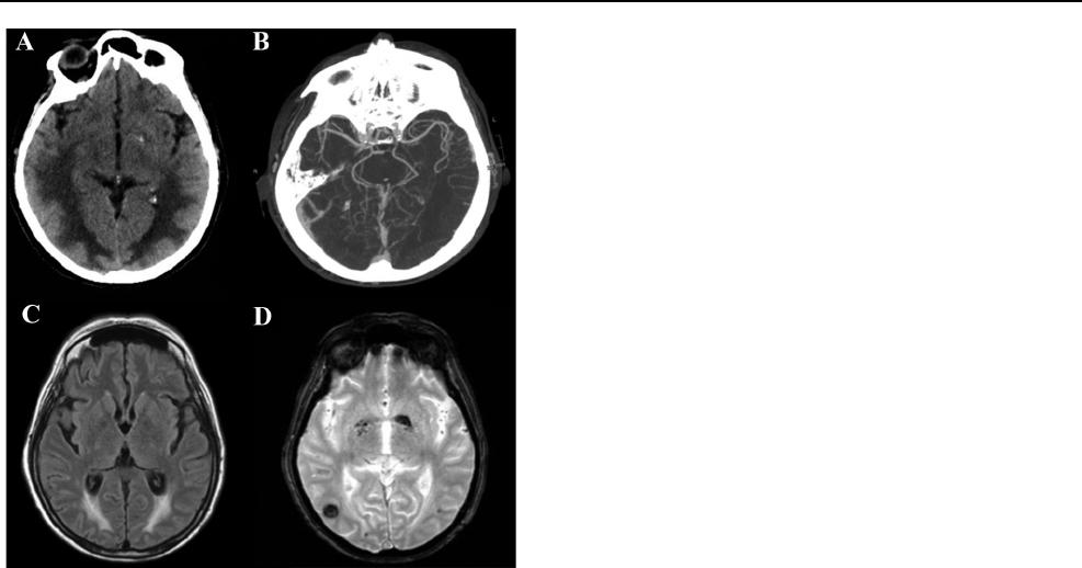
CT and CTA were consistent with hemorrhagic Posterior
Reversible Encephalopathy Syndrome (PRES; Fig.1a, b).
In the following days spontaneous breathing was restored.
No epileptic seizures were reported during hospitalization.
On day 56 a brain MRI showed a reduction of the bilateral
edema with bilateral occipital foci of subacute hemorrhage
(Fig.1c, d). A second nasopharyngeal swab was negative for
SARS-CoV-2, and she was alert and fully oriented with a nor-
malization of blurred vision.
Table 1 Laboratory and
neurophysiologic assessment
Assessment Exams
Laboratory At admission in Emergency Department
Vital Signs: blood pressure—150/70mmHg, heart rate 90 beats per minute, respira-
tory rate was 22 breaths per minute, oxygen saturation: 88% in air room
Arterial blood gas: pH 7.48, pCO
2
27.9mmHg, pO
2
82.2mmHg, lactates
1.56mmol/L, CHCO
3
24.5mmol/L
Blood count: Red cells: 4.11 10
12
/L (4–5.5), Hemoglobin: 12.1g/dL (12–16.5),
White cells: 7.21 10
9
/L (4.3–11), Neutrophils: 84%, Lymphocytes: 12%, Mono-
cytes: 4%, Platelets: 180 10^9/L (150–450)
Reactive C protein: 245.5mg/L (0–5), Creatinine: 1.20mg/dL, AST: 83 U/L (11–34),
ALT: 87 U/L (8–41), LDH:481 U/L (125–220), Glucose: 122mg/dL (74–109)
Infectious diseases
Day 0:
Real-Time PCR oropharyngeal swab SARS-CoV-2: positive
Day 2:
Mycoplasma, Legionella, Chlamydia Pneumoniae Antibodies/antigen: negative;
Day 12:
Urine culture:positive (Candida Albicans > 100,000CFU/mL) treated with flucona-
zole
Day 16:
Blood cultures: positive (St. Epidermidis) treated with piperacilin/tazobactam and
daptomicyn
Day 23:
Bronchial aspirate RNA SARS-CoV-2: negative
Urine culture: negative
Day 26:
Bronchial aspirate RNA SARS-CoV-2: negative
Day 29:
Blood cultures: positive (St. Epidermidis)
Day 33:
Real-Time PCR oropharyngeal swab SARS-CoV-2: negative
Day 44:
Mycoplasma, Legionella, Chlamydia Pneumoniae Antibodies/antigen: negative
Day 47:
Blood culture: negative
Autoimmune assessment
ANA: positive 1:160 homogeneous pattern; ANCA, (PR3)-Anti-Neutrophil Cytoplas-
mic Antibodies, MPO neutrophil antigen: negative
Immunological assessment
Lymphocyte typing: 918 cells/uL ( 1.000–4.000), CD3% antigen: 62% (60–86), anti-
gen CD3 573 cells/uL ( 836–2644), CD4% antigen:23 (30–60), CD4 antigen: 213
clls/uL:493–1772, CD8% antigen: 38%(16–42), CD4/CD8: 0.6 (1.0–2.2), CD16/
CD56% antigen: 2 (3–24), CD19% antigen: 34 (5–22)
CSF of lumbar puncture
Clear, colorless, normal pressure, glucose: 139mg/dl, protein: 53mg/dl, cells: 0.8
mm
3
; Microscopic examination: negative for
HSV 1–2 DNA, VZV DNA, Mycobacterium, Borrelia-Antibodies, COVID19 tested
on CSF: negative
Thyroid function: 1.140 McUI/mL (0.270—4.200)
Neurophysiology EEG: globally slow activity, with focus on the central-temporal and posterior regions
EMG/ENG: bilateral compressive common peroneal nerve axonal neuropathy
Journal of Neurology
1 3
Discussion
PRES is characterized by acute impairment in level of con-
sciousness, headache, visual disturbances and seizures, with
cortical/subcortical vasogenic edema, involving predomi-
nantly the parietal and occipital regions bilaterally [1]. PRES
is commonly associated with blood pressure fluctuations,
renal failure, autoimmune conditions, sepsis, preeclampsia
or eclampsia and immunosuppressive-cytotoxic drugs. In
our patient the sepsis (Table1) was due to Staph. Epider-
midis, that has never been associated with PRES, and did
not induce a shock condition as is usually the case in septic
PRES [2–4]. None of the drugs given to our patient has been
associated with PRES [5].
Several studies suggested a key role of endothelial dys-
function (ED), combined with hemodynamic stress (hyper-
tensive crisis) and immunological activation with release of
cytokines (TNF-α, IFN-γ, IL-1) able to activate endothelial
cells, thus increasing vascular permeability. ED is a prin-
cipal determinant of microvascular perfusion: by shifting
the vascular equilibrium towards a more pro-inflammatory,
pro-coagulant and proliferative state, it leads to ischaemia
and inflammation with edema [6].
This is the second report of hemorrhagic PRES in
COVID-19, and these other two patients were very similar
to ours.[7].
Mounting evidence suggests that the SARS-Cov2
directly infects endothelial cells causing diffuse inflamma-
tion [8–10]. The pivotal host cell receptor for the entry of
SARS-CoV-2 into the cells is the Angiotensin-Converting
Enzyme 2, which is also expressed by the brain endothe-
lium [9, 11]. Varga etal. [10] showed the presence of viral
elements within endothelial cells in different vascular beds,
suggesting a role of an ED in the systemic toxicity caused
by the virus.
In our patient we can rule out the causes of PRES listed
above. A contribution from the respiratory distress was
unlikely since PRES developed during mechanical ventila-
tion. We hypothesize that SARS-CoV-2 may have caused a
cerebrovascular ED which in turn was responsible for both
the hemorrhagic lesions and the for the disruption of the
blood brain barrier with vasogenic edema.
Availability ofdata andmaterial
Our data are available upon request to the corresponding
author.
Funding This research received no specific grant from any funding
agency in the public, commercial, or not-for-profit sectors.
Compliance with ethical standards
Conflicts of interest The authors declare no potential conflicts of inter-
est with respect to the research, authorship, and/or publication of this
article.
Consent for publication A written informed consent was obtained from
the patient.
Ethics approval Not applicable. This case has been described retro-
spectively, without the patient undergoing procedures and tests other
than those she already had to undergo to treat her clinical condition.
This research was performed in accordance with GCP and the ethical
standards laid down in the 1964 Declaration of Helsinki.
References
1. Hinchey J, Chaves C, Appignani B, Breen J, Pao L, Wang A, Pes-
sin MS, Lamy C, Mas JL, Caplan LR (1996) A reversible posterior
leukoencephalopathy syndrome. N Engl J Med 334(8):494–500.
https ://doi.org/10.1056/NEJM1 99602 22334 0803
2. Bartynski WS, Boardman JF, Zeigler ZR, Shadduck RK, Lister J
(2006) Posterior reversible encephalopathy syndrome in infection,
sepsis, and shock. AJNR Am J Neuroradiol 27(10):2179–2190
3. Racchiusa S, Mormina E, Ax A, Musumeci O, Longo M, Granata
F (2019) Posterior reversible encephalopathy syndrome (PRES)
Fig.1 Radiological findings. a Brain axial CT on day 25 shows pos-
terior frontal and temporo-parieto-occipital symmetric bilateral
hypodensity of the subcortical white matter, and a tiny left occipi-
tal parenchymal hemorrhage. b Para-axial CTA scan confirms the
absence of vascular malformation and alterations of posterior circle
vessel caliber, suggestive of vasoconstriction mechanism. c Axial T2
Flair image on day 56 shows that vasogenic edema is reduced but still
detectable and d T2 Gradient-Echo reveals the onset of right temporal
hypodensity, correlated to hemorrhagic process

Journal of Neurology
1 3
and infection: a systematic review of the literature. Neurol Sci
40(5):915–922. https ://doi.org/10.1007/s1007 2-018-3651-4
4. Toledano M, Fugate JE (2017) Posterior reversible encephalopa-
thy in the intensive care unit. Handb Clin Neurol 141:467–483.
https ://doi.org/10.1016/B978-0-444-63599 -0.00026 -0
5. Pilato F, Distefano M, Calandrelli R (2020) Posterior reversible
encephalopathy syndrome and reversible cerebral vasoconstriction
syndrome: clinical and radiological considerations. Front Neurol
11:34. https ://doi.org/10.3389/fneur .2020.00034
6. Fugate JE, Rabinstein AA (2015) Posterior reversible encepha-
lopathy syndrome: clinical and radiological manifestations, patho-
physiology, and outstanding questions. Lancet Neurol 14(9):914–
925. https ://doi.org/10.1016/S1474 -4422(15)00111 -8
7. Franceschi AM, Ahmed O, Giliberto L, Castillo M (2020) Hemor-
rhagic posterior reversible encephalopathy syndrome as a mani-
festation of COVID-19 infection. AJNR Am J Neuroradiol. https
://doi.org/10.3174/ajnr.A6595
8. Guo J, Huang Z, Lin L, Lv J (2020) Coronavirus disease 2019
(COVID-19) and cardiovascular disease: a viewpoint on the
potential influence of angiotensin-converting enzyme inhibitors/
angiotensin receptor blockers on onset and severity of severe acute
respiratory syndrome coronavirus 2 infection. J Am Heart Assoc
9(7):e016219. https ://doi.org/10.1161/JAHA.120.01621 9
9. Sardu C, Gambardella J, Morelli MB, Wang X, Marfella R, San-
tulli G (2020) Hypertension, thrombosis, kidney failure, and
diabetes: is COVID-19 an endothelial disease? A comprehensive
evaluation of clinical and basic evidence. J Clin Med. https ://doi.
org/10.3390/jcm90 51417
10. Varga Z, Flammer AJ, Steiger P, Haberecker M, Andermatt R,
Zinkernagel AS, Mehra MR, Schuepbach RA, Ruschitzka F, Moch
H (2020) Endothelial cell infection and endotheliitis in COVID-
19. Lancet 395(10234):1417–1418. https ://doi.org/10.1016/S0140
-6736(20)30937 -5
11. Natoli S, Oliveira V, Calabresi P, Maia LF, Pisani A (2020) Does
SARS-Cov-2 invade the brain? Translational lessons from animal
models. Eur J Neurol. https ://doi.org/10.1111/ene.14277
